Cataract Surgery
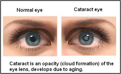
To comprehend cataracts, visualize the eye as a camera with a transparent lens focusing light on the reel of the eye, i.e. the retina. The created image on the retina is subsequently transferred to the brain via a nerve to complete the process of seeing. When this lens becomes opaque or white, the condition is known as a cataract. When specific proteins in the lens create aberrant clumps, the disease occurs. These clumps eventually expand and obstruct vision by distorting or preventing the passage of light through the lens. Dr. Himashree Wankhede is providing total Eye care Treatment in Wakad and she is the Best Ophthalmologist in Wakad.
When the lens is partially opaque, this is referred to as an infantile cataract, and some light can pass through to assist with everyday functions. As the opacity engulfs the entire lens, vision is completely lost, and the cataract is referred to as mature. Dr. Himashree Wankhede has been practicing Complete eye care therapy in Wakad for about 7 years. She is Top Ophthalmologist in Wakad.
Symptoms Of indicating Cataract
The vision gradually deteriorates without pain. An early cataract is connected with difficulty reading in normal light circumstances, necessitating the need for additional lighting. Night driving can be difficult due to glare and reduced clarity. Some people have fast variations in the number and power of their glasses. In extreme cases, there is full blindness and the lens turns pearly white in color.
If you encounter any of the following symptoms, make an appointment with your eye doctor every away:
- The vision that is cloudy or blurry
- Dual vision (diplopia)
- The fading of colors
- Noticing halo effects around lights
- heightened sensitivity to glare
- A type of vision distortion in which items appear to be seen through a veil.
What are the different types of cataract surgery?
The conventional cataract surgical technique is conducted in a hospital or an outpatient surgery center. The facility does not allow overnight stays. The most frequent type of cataract surgery today is termed phacoemulsification. After numbing the eye with drops or an injection, your surgeon will create a very small incision in the surface of the eye in or near the cornea using an operating microscope. A narrow ultrasound probe, which patients frequently confuse with a laser, is placed into the eye and employs high-ultrasonic vibrations to break apart the clouded lens (phaco emulsify). Using the same ultrasound probe, these minute shattered fragments are suctioned out of the eye. Phacoemulsification: As previously said, this is the most prevalent form of cataract removal. With this most recent method, cataract surgery can usually be finished in less than 30 minutes with minimal sedation. Numbing eye drops or an injection around the eye are utilized, and no stitches or eye patches are usually required after surgery. Although lasers are not utilized in phacoemulsification, a femtosecond laser may be used to open the anterior capsule of the lens just prior to emulsification.
Pterygium
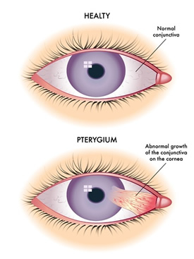
It is a degenerative disorder of the subconjunctival tissue, which proliferates as a triangular fold of tissue mass to invade the cornea, affecting the Bowman’s membrane and the superficial stroma, and is completely covered by conjunctival epithelium. It literally means “a wing”.
It is unclear what causes a pterygium to form. Nonetheless, most experts agree that the following are major risk factors:
- Extensive UV light exposure
- A Dry Eye
- Dust and wind are irritants
Pterygium symptoms are determined by the clinical appearance of the lesion. Typical discoveries include:
- Fibrovascular conjunctival growth extending onto the corneal surface from the palpebral fissure
- Vascular straightening in the direction of the approaching pterygium's head of the pterygium on the corneal surface.
- May be a thin translucent membrane or significantly thickened with an elevated mound of gelatinous material.
- It may affect the nasal and temporal limbus of both eyes or only a single location.
- Raised lesion, white to pink in color depending on vascularity.
Treatment :
Today a variety of options are available for the management of pterygium, from irradiation to conjunctival auto-grafting or amniotic membrane transplantation, along with glue and suture application. As it is a benign growth, pterygium typically does not require surgery unless it grows to such an extent that it covers the pupil, obstructing vision or presents with acute symptoms. Some of the irritating symptoms can be addressed with artificial tears.
Glaucoma
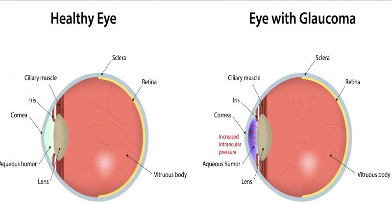
Glaucoma is a disease characterised by usually high pressure inside the eye that damages the optic nerve and may lead to permanent loss of vision. Not all 3 criteria (that is, high pressure inside the eye, optic nerve damage, and vision loss) are required to diagnose glaucoma; however, a diagnosis of glaucoma is certain when all 3 criteria are present.
What are the types of Glaucoma?
-
There are 2 main types of glaucoma:
- Open-Angle Glaucoma
- Angle Closure Glaucoma
What are the different types of cataract surgery?
Open-angle type of glaucoma usually does not give rise to any symptoms in early stages. In late stages, patients may feel pain in eyes and discomfort, and some individuals may notice field defects (inability to see certain areas of the field of vision). Usually, this type of glaucoma is diagnosed on examination by an eye specialist either when he suspects it because of some risk factors or during the course during the course of a routine examination.
Angle-closure type of glaucoma can give rise to pain in the eye and headache with vomiting, seeing coloured rings (haloes) around lights and redness in the eyes usually after coming out of a movie theatre. This type of glaucoma may occur as sudden attacks where there is severe pain in the eye, redness, watering, vomiting and blurring of vision.Treatment :
The treatment options for glaucoma include:
- Drugs
- Laser
- Operation
The treatment is decided by many factors:
- Type of glaucoma
- Stage of glaucoma
- Damage done
- Status of the other eye
- Response to other treatment already taken
- Patient compliance or reliability about taking drugs and follow up examination.
The decision regarding what treatment and when to be used should be left to the judgment of consulting eye surgeon.
Detected early and treated properly, glaucoma is perfectly compatible with lifelong good vision. If neglected it can end in blindness.
Keratoconus
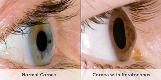
Keratoconus usually starts in the teenage years. It can, though, begin in childhood or in people up to about age 30. It’s possible it can occur in people 40 and older, but that is less common.
The changes in the shape of the cornea can happen quickly or may occur over several years. The changes can result in blurred vision, glare and halosat night, and the streaking of lights.
The changes can stop at any time, or they can continue for decades. There is no way to predict how it will progress. In most people who have keratoconus, both eyes are eventually affected, although not always to the same extent. It usually develops in one eye first and then later in the other eye.
With severe keratoconus, the stretched collagen fibres can lead to severe scarring. If the back of the cornea tears, it can swell and take many months for the swelling to go away. This often causes a large corneal scar.
Our eye surgery services include cataract removal, retinal procedures, and other advanced surgical treatments to improve your vision.
Keratoconus Treatment
Wankhede Eye Hospital takes pride in being one of the most widely recognized eye hospitals in Pune offering best services in Keratoconus treatment through continual improvements in our systems, technology and dedicated professionals. We are committed to offering new hope and solutions to all our patients at affordable cost.
Keratoconus is a progressive disease in which the patient’s cornea that is usually round in shape becomes thin and irregular shaped (cone-like). Due to this, the light rays entering the eye don’t focus on a single point resulting in blurred and distorted vision. If left untreated, Keratoconus becomes severe, leading to sudden & significant vision loss. Generally, it affects both eyes differently and its initial symptoms can be most commonly found in late teens. Wankhede Eye Hospital is well-equipped with the latest technology and ultra-modern equipment for the early diagnosis and monitoring of Keratoconus. We have an extremely talented, skilled and experienced team of eye surgeons in Pune offering a wide spectrum of Keratoconus treatments that include:
- Corneal collagen cross-linking(CXL)
- Contact Lenses (Custom Soft, Gas Permeable, Piggybacking, Hybrid, Scleral and Semi-scleral)
- Topography-guidedConductive Keratoplasty
- Corneal Transplant
In the initial stages of Keratoconus, CXL has proved to be very successful in correcting the blurred and distorted vision. But in advanced stages, when CXL and other treatments don’t provide acceptable vision, then corneal transplant is considered to get required outcomes. Our expert Keratoconus specialists perform cornea transplant procedure with high accuracy & precision. They replace the diseased cornea with a healthy one using advanced techniques and give the most successful results.
Lasik Surgery
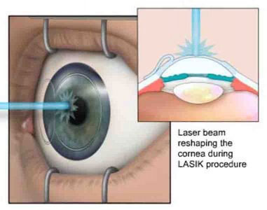
LASIK, which stands for laser in-situ keratomileusis, is a popular surgery used to correct vision in people who are nearsighted, farsighted, or have astigmatism.
All laser vision correction surgeries work by reshaping the cornea, the clear front part of the eye, so that light traveling through it is properly focused onto the retina located in the back of the eye. LASIK is one of a number of different surgical techniques used to reshape the cornea.
What Are the Advantages of Lasik Surgery? :
- It works! It corrects vision. Around 96% of patients will have their desired vision after LASIK. An enhancement can further increase this number.
- Vision is corrected nearly immediately or by the day after LASIK.
- No bandages or stitches are required after LASIK.
- Adjustments can be made years after LASIK to further correct vision if vision changes while you age.
- After having LASIK, most patients have a dramatic reduction in eyeglass or contact lens dependence and many patients no longer need them at all.
Femtosecond Laser
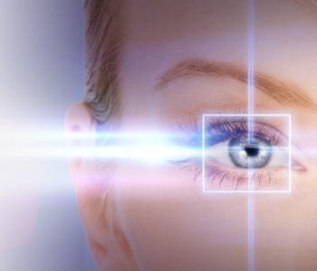
Bladeless Lasik involves the use of two lasers – first is the Femtosecond Laser, which is used to create an ultra-thin flap in the cornea, second is Excimer Laser which is used to reshape the cornea to induce correction. The flap is then positioned back which sticks to its place immediately. No blades are used at any time in the whole procedure.
Advantages of Bladeless Lasik :
- No fear of blades working on an eye.
- Precise cutting resulting in better visual outcomes and faster recovery.
- Ultra-thin and better quality flap – Safer than Blade Lasik.
- More customized treatment for patients
- Tissue Saving
- Faster Recovery
Dry Eye
What is Dry Eye?
Tears Keep the surface of the eyes wet, clean and protect your eyes from infections Tears also provide constant moisture and lubrication to maintain the vision and comfort to the eyes. If your eyes do not produce enough tears or if the tears dry up too quickly, the condition is termed as dry eye.
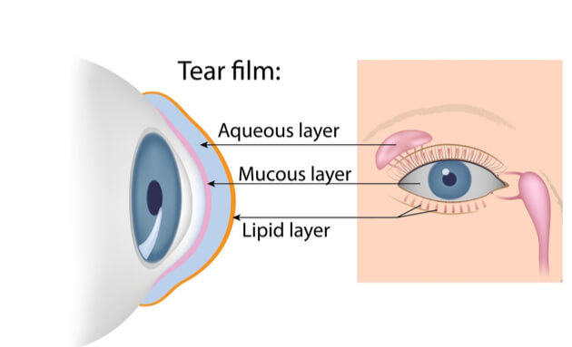
What Causes Dry Eye?
Dry eye may be temporary or a chronic condition.
Different causes for dry eye are as follow:
- Dry, dusty or windy climate, air conditioning also can result in increased tear evaporation resulting in dry eye.
- Long-term use of contact lens can also result in dry eye.
- Infrequent blinking associated with staring at the computer or video screens may also lead to dry eye symptoms Skin disease on or around the eyelids can result in dry eye.
- Dry eye can also develop after the refractive surgery knows as LASIK. However, these symptoms generally last 3-6 months but may last longer in some cases
- Diseases of the glands in the eyelids can cause dry eye.
What is the treatment for Dry Eye?
Numerous treatment strategies exist to battle the signs and symptoms associated with dry eye. Some of them include:
- The blinking exercise of the eye.
- Cleansing the eyelids with an application of warm compress.
- Using artificial tears, gels, lubricating drops and Ointments.
- Avoiding smoke, dust, air-conditioners and heaters.
- Increasing humidity in your surrounding by using a dish containing water on a window sill or radiator.
- Plugging the drainage holes, small circular openings at the inner corners of the eyelids where tears drain from the eye into the nose.
- Increasing water intake.
- Avoiding alcohol and caffeine.
- Increasing diet intake rich in polyunsaturated fatty acids as it reduces ocular surface inflammation and improves dry eye symptoms.
- Lowering the computer screen to below eye level.
- Taking regular breaks and blinking frequently during sessions in front of the computer.
Retina
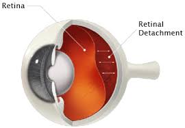
A Detached Retina is a serious and sight-threatening event. It occurs when the retina becomes separated from its underlying supportive tissue. The retina cannot function when these layers are detached. And unless the retina is reattached soon, it may result into permanent loss of vision.
Retinal Detachment could be a result of an eye or face injury, cataract surgery, tumors, or eye diseases. But diabetes may also cause Retinal Detachment. It usually occurs when a tear or hole in the retina allows the liquid vitreous to get under the retina and accumulate. The retina separates from the back wall of the eye. This detachment causes symptoms of a gradually enlarging veil or dark shadow in the peripheral visual field. When the detachment extends into the central part of the retina (macula) the central vision is lost.
Retinal Detachment should be fixed immediately through a retinal detachment surgery by a retina specialist to prevent complete loss of vision. Ideally, the Retinal Detachment should be repaired prior to the involvement of the macula. If the macula becomes detached, the vision is unlikely to be as good as it was prior to the Retinal Detachment even with successful surgical reattachment.
How can diabetes affect eye? You may have heard that diabetes causes eye problems and may lead to blindness. People with diabetes do have a higher risk of blindnessand other various eye problems than people without diabetes. Diabetes can affect the eyes and vision in a number of ways. It may lead to frequent fluctuations in vision, cataract in young age, decreased vision due to involvement of optic nerve, temporary paralysis of the muscles controlling the movement of eyes and thus double vision, the Incidence glaucoma is higher in diabetics. The most significant complication of diabetes in eye is diabetic retinopathy and its complications.
Symptoms :
- When detachment occurs, appearance of curtain falling in front of the eye, decreased vision or total obscuration of vision may happen.
- Bright flashes of light, especially in peripheral vision is felt
- Blurred vision
- Floaters in the eye
- Sudden appearance of shadow or blindness in a part of the visual field of one eye is experienced
Age Related Macular Degeneration (ARMD):
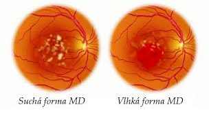
- The painless disorder that affects the macula, in one or usually both eyes, causing progressive loss of central and detailed vision.
- It is the leading cause of significant vision loss among patients over the age of 50.
- Does not lead to total blindness but patient may find it difficult to do read, drive and recognize people.
- The area surrounding the macula is not affected so peripheral vision remains clear and patient can usually still move around fairly freely.
Diagnosis And Treatment:
The diagnosis of ARMD is conducted through the series of latest and advanced tests. We provide the best and cost-effective treatments for age-related macular degeneration. In many cases, the risk of progression of visual loss caused due to Dry ARMD can be decreased by giving the high dose of multivitamins. But for the Wet form of ARMD, there are a few treatment options available, like intravitreal injections, lasers. However, in both types of ARMD, the best outcomes occur if the disease is diagnosed before much damage is caused to eyes. In patients with severe visual loss LOW VISUAL AIDS can be prescribed for improving the quality of vision enabling him/her to carry on their daily routine.

Pediatric Eye Checkup
Pediatric Ophthalmology is a subspecialty of ophthalmology dealing with eye problems in children. Clear vision plays an important role in the mental, physical, and social development of children. Amblyopia, or “lazy eye,” is a common eye problem in children. The problem occurs when the pathways of vision in the brain don’t develop properly. The human visual system develops as the brain matures; this is a process that takes about ten years. It is important to diagnose and treat amblyopia at an early age. If not caught early enough, children with amblyopia have a risk of permanent vision loss.
Diabetic Eye Disease
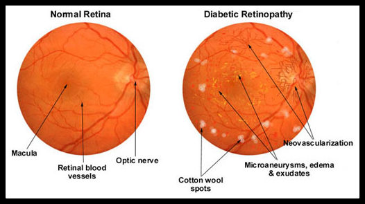
Diabetes and eye diseases go hand in hand. As a matter of fact, one of the major concerns for diabetic patients is that they might lose their eyesight if their sugar levels are not kept under control for long. A question raging in the minds of people might be how can diabetes which is a disease of the pancreas can affect such a dire effect on your vision? This can be simply explained by the occurrence of diabetes in our bloodstream. When we eat food, it gives us glucose which when broken by the natural insulin of our body releases small cells of energy. In a diabetic patient, his natural insulin mechanism fails and the unbroken glucose gets absorbed into our bloodstream thus causing elevated levels of sugar in our body. You may have heard that diabetes causes eye problems and may lead to blindness. People with diabetes do have a higher risk of permanent blindness and other various eye problems than people without diabetes. Diabetes can affect the eyes and vision in a number of ways. It may lead to frequent fluctuations in vision, cataract in young age, decreased vision due to an involvement of optic nerve, temporary paralysis of the muscles controlling the movement of eyes and thus double vision, the Incidence glaucoma is higher in diabetics. The most significant complication of diabetes in an eye is diabetic retinopathy. This may cause irreversible loss of vision in later stages. Visionnex Eye Center we not only provide excellent medical facility and advanced technology to deal with diabetic eye diseases but also help restore your vision to a larger extent if not fully.
Implantable Contact Lense

Our vision correction services include a range of treatments to correct refractive errors, helping you achieve clear and sharp vision.
WHAT IS THE VISIAN ICL?
The Visian ICL is a special contact lens that is implanted inside the eye and works with the eye’s natural lens to produce sharp, clear vision. Crafted from Collamer, an advanced biocompatible material, the Visian ICL Implantable Contact Lens is completely safe and long-lasting. Our ICL experts find that the Visian ICL greatly increases the number of patients who are able to reduce or even eliminate their dependence on glasses or traditional contact lenses.
Good candidates for the ICL include patients who:?
- Are between the ages of 18 and 45 years.
- Have dry eyes, very high myopia (above -12.00D), or a thin cornea (non-LASIK candidate).
- Are nearsighted or farsighted, including those with mild, moderate, and high power with or without occurrence of astigmatism. The Toric ICL, capable of correcting myopia and astigmatism together and combines two procedures into one.
- Have proper anterior chamber depth as will be determined by the eye surgeon or ophthalmologist after a comprehensive eye exam.
- Have not had a change in their eyeglass prescription of more than 0.50 Diopters in a year.
- Are not currently pregnant.
- Have no known allergies to medications used during refractive surgery or no other contraindications.
The ICL is made of Collamer, a highly biocompatible advanced lens material which contains a small amount of purified collagen. Collamer does not cause a reaction inside the eye and it contains an ultraviolet filter that provides protection to the eye.
Currently we use ICL’s from the STAAR Surgical Company, the company that manufactures ICL. The Toric ICL is only a variant which corrects your nearsightedness as well as your astigmatism (cylindrical power) in one single procedure.
All ICL’s are custom made to meet the needs of each individual eye, after measurements and data are taken and sent to the manufacturing company. This type of lens implant has been available internationally for over 12 years. The implant does not treat presbyopia (difficulty with reading in people 40 and older), but you can use reading glasses as needed after receiving the ICL.
Contact lens
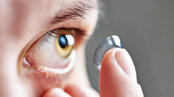
A contact lens, or simply contact, is a thin lens placed directly on the surface of the eye. Contact lenses are considered medical devices and can be worn to correct vision, or for cosmetic or therapeutic reasons When compared with spectacles, contact lenses typically provide better peripheral vision, and do not collect moisture such as rain, snow, condensation, or sweat. This makes them ideal for sports and other outdoor activities. Contact lens wearers can also wear sunglasses, goggles, or other eyewear of their choice without having to fit them with prescription lenses or worry about compatibility with glasses. Additionally, there are conditions such as keratoconus in which contact lens helps a lot in getting better vision.
What are the types of Contact Lenses available?
There are basically two types of contact lenses:
1. Rigid Gas-permeable (RGP) Contact Lenses which are also known as “semi-soft lenses”.
2. Soft Contact Lenses Hard Contact Lenses have become obsolete now.
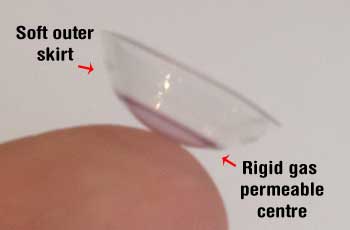
Soft lenses can be further classified depending on the type of wear:
- Daily wear
- Extended wear
- Disposable (Quarterly, Monthly, Fortnightly, Weekly and Daily)
Services We Provided
- Dispensing of different types of regular contact lenses – daily wear and extended wear Soft Disposable Lenses, Soft Conventional Lenses, Rigid Gas Permeable Lenses, Cosmetic lenses, Prosthetic lenses, and Bandage contact lenses.
- Dispensing Specialty Contact Lenses – Scleral Contact Lenses, Soft Toric Lenses, Rose-K Lenses for Keratoconus, Bifocal Contact Lenses, Piggyback Lenses for Keratoconus, Soft-perm lenses.
- Dispensing of appropriate Contact Lens Solutions for different type of lenses
